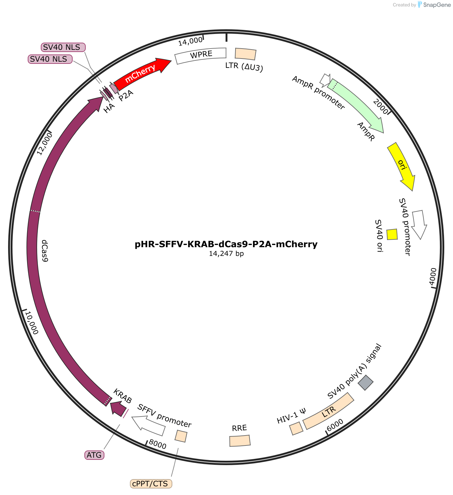

Therefore, up to 50% of F2A-linked proteins can remain in the cell as a fusion protein, which might cause some unpredictable outcomes, including a gain of function. Incomplete cleavage ĭifferent 2A peptides have different efficiencies of self-cleaving, T2A and P2A being the most and F2A the least efficient. ĢA peptides, when combined (or not) with the IRES elements, can make it possible to generate multiple separated peptides within a single transcript. Inserting the CDS of a 2A peptide into the fusing point or replacing the linker sequence with the CDS of a 2A peptide protein can cleave the fused protein into two separated peptides, rescuing the function of the two peptides. The 2A peptides can be applied when the fused protein doesn’t work. In genetic engineering, the 2A peptides are used to cleave a longer peptide into two shorter peptides. However, it is believed to involve ribosomal "skipping" of glycyl-prolyl peptide bond formation rather than true proteolytic cleavage. The exact molecular mechanism of 2A-peptide-mediated cleavage is still unknown.

The apparent cleavage is triggered by ribosomal skipping of the peptide bond between the Proline (P) and Glycine (G) in C-terminal of 2A peptide, resulting in the peptide located upstream of the 2A peptide to have extra amino acids on its C-terminal end while the peptide located downstream the 2A peptide will have an extra Proline on its N-terminal end. Adding the optional linker “ G S G” (Gly-Ser-Gly) on the N-terminal of a 2A peptide helps with efficiency.

The following table shows the sequences of four members of 2A peptides. F2A is derived from foot-and-mouth disease virus 18 E2A is derived from equine rhinitis A virus P2A is derived from porcine teschovirus-1 2A T2A is derived from thosea asigna virus 2A. Four members of 2A peptides family are frequently used in life science research.


 0 kommentar(er)
